Digital radiography is the modern method of taking dental X-rays using electronic sensors and computer software instead of traditional film. For patients, this translates to faster appointments and clearer images that your dental team can examine together in real time. The technology supports more informed decision-making by giving clinicians immediate access to high-quality visuals of the teeth, roots, and surrounding bone.
One of the most important advantages is a significant decrease in radiation exposure compared with older film techniques. Advances in sensor sensitivity and image-processing software mean we can capture diagnostically useful images using lower doses, which is particularly reassuring for patients who require routine monitoring or those who are pregnant or medically vulnerable.
Beyond safety, digital radiography integrates seamlessly with modern clinical workflows. Images are stored in the patient’s electronic record, allowing dentists and hygienists to track changes over time, compare past and present images, and create a clear visual record that supports long-term oral health planning.
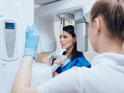
At the heart of digital radiography is a compact electronic sensor that temporarily sits in the patient’s mouth to capture an image. When the X-ray exposure occurs, the sensor converts the X-ray energy into an electrical signal, which is transmitted to a computer and rendered as an image in seconds. This immediate availability eliminates wait times associated with film development and lets clinicians review results with patients right away.
Once the image appears on screen, software tools can enhance it—adjusting contrast, magnifying areas of interest, or measuring distances with precision. These non-destructive edits assist clinicians in highlighting potential issues such as decay between teeth, root abnormalities, or early-stage bone loss without retaking images.
Storing images digitally also streamlines record keeping. Every radiograph is tagged to the patient’s file and can be retrieved instantly during subsequent visits. This continuity helps the dental team detect subtle changes over months or years, supporting preventive care and early intervention.
Digital radiography sharpens the detection of dental problems at an earlier stage than many physical exams alone. Enhanced imaging reveals minute details—tiny cavities beneath fillings, hairline fractures, and early signs of periodontal disease—that might otherwise go unnoticed. Early detection often makes treatments less invasive and more predictable for patients.
Because images can be manipulated for clarity, clinicians can better visualize the anatomy of tooth roots and surrounding bone. This level of detail is particularly valuable when planning endodontic procedures, implant placement, or evaluating the fit and integrity of restorations. Precise images reduce guesswork and help tailor treatments to each patient’s unique anatomy.
Another practical benefit is improved interdisciplinary communication. Digital files can be exported or shared with specialists when consultation is needed, allowing for collaborative treatment planning without redundant imaging. That coordination helps streamline care and ensures everyone involved has the same accurate information.
The process of having digital radiographs taken is quick and straightforward. A small sensor is placed inside the mouth while the patient bites down briefly; the exposure lasts only a fraction of a second. Most patients report minimal discomfort—often less than traditional film—because the sensors are slimmer and the workflow is faster.
Safety protocols are always followed to minimize exposure. Lead aprons and thyroid collars may be used when appropriate, and exposures are kept as low as reasonably achievable. For patients with special needs, adjustments to positioning and technique are routinely made to ensure comfort and accuracy in imaging.
After the images are captured, your dental provider will review them with you on a chairside monitor. This visual review is an opportunity to ask questions and better understand any recommended next steps. Patients often find it reassuring to see the actual images and to have their clinician explain what the visuals mean for their oral health.
Digital radiography is not only clinically superior but also more environmentally friendly than film-based systems. Because no chemical developers or paper processing are required, the practice eliminates harmful waste associated with darkroom processing. This reduces the clinic’s environmental footprint while simplifying office logistics.
From a records perspective, digital files are encrypted and stored within the practice’s secure electronic health record system. This approach supports both long-term archival and quick retrieval while adhering to privacy and data-protection standards. When appropriate, images can be exported for referral or second opinions, preserving quality without physical transport.
The ability to instantly share and review images also reduces the need for repeat exposures. When collaborating with outside specialists or transferring records to another provider, digital files arrive intact and diagnostically useful, often avoiding unnecessary retakes that would expose patients to additional radiation.
Digital radiography represents a meaningful improvement in dental imaging—safer, faster, and more informative than film-based X-rays. By delivering immediate, high-resolution images and simplifying storage and sharing, the technology supports more accurate diagnoses and better-informed treatment planning. Patients benefit from reduced radiation exposure, shorter appointment times, and clearer communication with their dental team.
At Wells Dentistry, we use digital radiography as part of a broader commitment to modern, patient-centered care. If you have questions about what to expect or how digital imaging fits into your treatment plan, please contact us for more information.


Digital radiography is a method of taking dental X-rays that uses electronic sensors and computer software to create images instead of traditional film. The sensor captures X-ray energy and converts it into a digital file that appears on a monitor within seconds. This immediate visualization helps clinicians and patients review images together during the same appointment.
Because images are produced digitally, they can be enhanced, measured, and archived without chemical processing or physical storage. The workflow is faster and more efficient, allowing dental teams to compare current images with prior records to monitor changes over time. Digital files can also be shared securely with specialists when collaborative care is needed.
Digital radiography eliminates darkroom processing and film handling by creating images electronically, which reduces wait times and environmental waste. Sensors are generally more sensitive than film, so diagnostically useful images can often be acquired with lower radiation doses. Software tools allow clinicians to adjust contrast, magnify areas of interest, and apply measurements without altering the original image.
Because images are immediately available on-screen, clinicians can confirm diagnostic quality during the appointment and retake images only when necessary, improving efficiency. Digital files are easier to organize within an electronic health record, which simplifies comparisons and long-term tracking. The ability to export images in standard formats supports referrals without needing physical film copies.
Digital radiography typically uses less radiation than conventional film techniques because sensors are more sensitive and image-processing software can enhance images captured at lower doses. Safety protocols such as collimation, proper positioning, and the use of lead aprons or thyroid collars further reduce exposure. Dental teams follow the ALARA principle—keeping radiation As Low As Reasonably Achievable—when selecting imaging settings and frequency.
For patients who require routine monitoring or have special medical considerations, clinicians tailor imaging schedules and techniques to balance diagnostic needs with safety. Pregnant patients are evaluated carefully, and imaging is performed only when necessary with appropriate shielding and justification. When used appropriately, digital radiography is a safe and effective diagnostic tool.
The process is quick and generally comfortable: a small sensor is positioned in the mouth while the patient bites briefly and a fraction-of-a-second exposure is made. Because sensors are thin and the workflow is immediate, many patients find digital imaging less intrusive and faster than film-based methods. Technicians take care to position sensors and the patient to capture diagnostically useful images with minimal repeats.
After images are taken, the clinician reviews them on a chairside monitor and explains any findings in real time. This is an opportunity to ask questions and see the visuals that support recommendations for treatment or monitoring. If additional views are needed, adjustments are made promptly so the appointment can proceed efficiently.
Digital images reveal fine details that may not be visible during a visual exam, such as small cavities between teeth, early bone changes from periodontal disease, hairline cracks, and root abnormalities. Software tools can enhance contrast and magnify specific areas to help clinicians detect subtle issues earlier. Early detection often allows for less invasive and more predictable treatment options.
Images are also used to document baseline conditions and follow progression over time, which is important for monitoring chronic conditions or the success of prior treatments. When planning restorative work, clinicians reference radiographs to assess tooth structure, root anatomy, and surrounding bone. Accurate imaging reduces uncertainty and helps tailor treatments to each patient’s anatomy.
Yes, digital radiographs are essential tools for planning many advanced dental procedures because they provide clear views of tooth roots, surrounding bone, and adjacent anatomical structures. High-resolution intraoral images help assess root morphology and detect periapical pathology relevant to endodontic treatment. For implant planning, two-dimensional digital images are often combined with other imaging modalities to evaluate bone height and spacing precisely.
When more detailed three-dimensional information is required, clinicians may recommend cone-beam computed tomography (CBCT) in addition to standard digital radiographs to measure bone volume and avoid critical structures. Using high-quality images reduces guesswork, supporting safer, more predictable outcomes. At Wells Dentistry, our clinical team integrates digital imaging into treatment planning to enhance precision and patient understanding.
Digital radiographs are stored electronically within the practice’s secure patient record system and are typically encrypted and backed up according to healthcare privacy standards. Files are tagged to each patient’s chart and can be retrieved instantly during follow-up appointments, which streamlines continuity of care. Secure storage also facilitates the controlled sharing of images when specialist consultation or a referral is necessary.
Strict access controls and routine backups reduce the risk of data loss and help maintain confidentiality. Because there is no chemical processing, digital workflows also eliminate hazardous waste associated with film development. Patients can request copies of their images for transfer to other providers when needed, and electronic formats preserve diagnostic quality during exchange.
Yes, clinicians adapt positioning, sensor selection, and exposure settings to suit children and patients with special needs to ensure diagnostic quality while maximizing comfort. Pediatric sensors and smaller exposure fields help reduce dose and make the process less intrusive for younger patients. Technicians may take extra time to explain procedures, use gentle positioning techniques, and employ behavior management strategies as needed.
For patients who have difficulty tolerating intraoral sensors, alternative projections or modified approaches can often provide adequate diagnostic information. The dental team balances the need for diagnostic imaging with the patient’s comfort and ability to cooperate, always following safety protocols to minimize exposure. Tailoring imaging protocols helps ensure that all patients receive appropriate and compassionate care.
Common digital imaging modalities include intraoral sensors for bitewing and periapical images, panoramic radiography for comprehensive arch views, and cone-beam computed tomography (CBCT) for three-dimensional assessment when detailed spatial information is required. Each modality serves a different diagnostic purpose: intraoral images capture fine tooth detail, panoramic images show broader structures, and CBCT reveals volumetric bone and anatomical relationships. Clinicians select the modality that answers the clinical question while minimizing unnecessary exposure.
Advances in software integration allow images from these modalities to be viewed, measured, and annotated in a unified workspace, improving diagnostic clarity and treatment planning. The availability of multiple imaging options supports personalized care tailored to each patient’s needs. Appropriate selection and combination of modalities enhance diagnostic confidence and procedural outcomes.
Digital radiography enhances patient communication by enabling clinicians to display and explain images during the appointment, which helps patients understand diagnoses and treatment options more clearly. Visuals that can be annotated or magnified make complex findings easier to grasp and support informed decision-making. This transparent review of images builds trust and allows patients to participate actively in their care planning.
From a continuity standpoint, digital files are simple to archive and retrieve, so changes can be tracked across visits and shared with specialists without loss of quality. Electronic sharing reduces the need for repeat exposures and fosters coordinated treatment among multiple providers. By improving clarity and recordkeeping, digital imaging supports safer, more efficient care over the long term, and it is a standard tool used across modern dental practices including Wells Dentistry.
It's time for your check-up!
Scheduling your next visit or getting answers about our comprehensive dental services is simple when you contact Wells Dentistry. Our friendly administrative team is prepared to help you find the perfect appointment time, provide clarity on your treatment plan options, and efficiently manage any billing or insurance questions. We are ready to assist you by phone or via our quick online form. Contact us today and let us partner with you to maintain a healthy, beautiful smile for years to come.