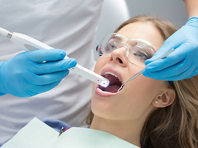
An intraoral camera is a compact, pen-sized imaging tool that captures high-resolution color photos of the teeth, gums, and other tissues inside the mouth. Designed to fit comfortably within the oral cavity, these devices provide a close-up view that goes far beyond what the naked eye can discern. The resulting images offer crisp detail of enamel, restorative materials, cracks, tartar, and soft-tissue changes that can be difficult to document with standard visual inspection alone.
Because intraoral cameras produce real-time images, clinicians can magnify trouble spots, compare surfaces from different angles, and pause on frames that reveal subtle signs of wear or early disease. This visual feedback is especially useful for monitoring progression over time: images taken at separate visits can be compared side-by-side to show improvement, stability, or the need for further evaluation. The photographic record also helps ensure nothing is overlooked during a clinical exam.
These cameras are carefully engineered for oral use, with smooth surfaces and ergonomic shapes that minimize discomfort. Many models incorporate LED lighting and anti-fog features to maintain image clarity, even in darker corners of the mouth. The result is a reliable, repeatable way to document oral health during routine checkups or focused diagnostic visits.
High-quality intraoral photos are more than just illustrations — they are clinical tools that enhance diagnostic accuracy. When a tooth surface looks suspicious under magnification, the camera view can help differentiate between staining, early decay, hairline fractures, or defective restorations. This level of detail allows clinicians to make better-informed decisions about follow-up imaging, preventive measures, or when to recommend restorative care.
Beyond identifying specific problems, intraoral images support more precise treatment planning. For example, clear photos of margins around crowns or fillings help determine whether a restoration is sealing properly, while detailed images of gum tissue can reveal early signs of inflammation or recession. These insights allow the dental team to tailor care to the patient’s needs and to sequence interventions more effectively.
Moreover, intraoral imaging integrates smoothly with other diagnostic technologies such as digital radiography and intraoral scanners. Together, these tools create a multi-dimensional record that strengthens clinical reasoning. By combining visual photos with x-rays and digital impressions, the care team builds a fuller understanding of both surface and internal conditions — improving outcomes and reducing the likelihood of surprises during treatment.
An intraoral camera exam is typically quick, comfortable, and noninvasive. During a routine checkup or targeted assessment, your dentist or hygienist will gently guide the camera into the mouth and capture a series of images. The clinician may ask you to reposition slightly so they can photograph different angles, but the process rarely causes discomfort and often takes only a few minutes within the course of an appointment.
As images are captured, they are displayed on a monitor so both clinician and patient can view them together. This immediate visual feedback helps explain findings in plain terms and allows for an interactive conversation about oral health. If an image reveals an area that requires attention, the clinician will describe the concern, explain recommended next steps, and document the observation in your record.
Images from the session are saved to the patient’s digital chart and can be reviewed later as part of ongoing care. Because the process is fast and repeatable, intraoral photography is suitable for both single-visit diagnostics and longer-term tracking of conditions like wear, staining, or tissue changes.
One of the most immediate benefits of intraoral imaging is improved communication. Seeing a magnified, full-color image of a problem area helps patients understand what the clinician sees and why a particular recommendation is being made. This clarity often reduces uncertainty and enables patients to make more informed decisions about their care.
Intraoral photos also support patient education. Clinicians can point out signs of early decay, plaque accumulation, or gum irritation and demonstrate how targeted home care or professional interventions can address those issues. Visual examples make it easier to discuss prevention strategies, such as improved brushing techniques, interdental cleaning, or scheduling regular professional cleanings.
Finally, saved images provide a transparent record of your oral health history. When it’s time to coordinate care with a specialist, laboratory, or other provider, those same images can be shared (with consent) to clarify treatment needs without relying solely on memory or descriptive notes. The result is a smoother, more collaborative experience for patients who want to be active participants in their care.
Intraoral images are typically stored digitally alongside radiographs, periodontal charts, and treatment notes, creating a centralized and searchable patient record. This digital integration simplifies follow-up appointments and makes it easier for clinicians to track changes over time. Having a visual archive reduces ambiguity and supports consistent, evidence-based decision-making during subsequent visits.
These images are also valuable when coordinating care. Whether a patient needs a referral for specialized treatment or restorative work that involves a dental laboratory, clear intraoral photos provide precise visual context. Labs can use these images to match shades, contours, and margins more accurately, while specialists receive a clear preview of the clinical situation before an in-person consultation.
At practices like Wells Dentistry, intraoral imaging is part of a broader commitment to leveraging modern technology for better patient outcomes. When used responsibly as part of a comprehensive diagnostic protocol, these images improve the efficiency of care, enhance documentation quality, and promote clearer communication among the dental team and patients.
In summary, intraoral cameras bring a new level of visibility to routine dental care: they reveal subtle problems, support more accurate diagnoses, and foster clearer communication between clinicians and patients. If you’d like to learn how intraoral imaging is used during exams or how it may benefit your specific dental needs, please contact us for more information.


An intraoral camera is a compact, pen-sized imaging device designed to capture high-resolution, full-color photos of the teeth, gums and oral tissues. The camera is inserted into the mouth and positioned near areas of interest while built-in lighting and optics produce clear images that magnify small details. These images are displayed in real time on a monitor and saved digitally to a patient record for later review.
The device works by combining LED illumination with a small sensor and lens system that delivers magnified views of tooth surfaces and soft tissue. Anti-fog and ergonomic housings keep the camera usable in tight or moist areas of the mouth and maintain patient comfort. Because the images are digital, clinicians can enlarge, compare and annotate frames to support diagnosis and communication.
Intraoral camera images often reveal subtle changes that are difficult to detect with the naked eye, such as hairline fractures, early enamel breakdown, marginal gaps around restorations or initial areas of discoloration. Magnified, well-lit photos make it easier to distinguish true pathology from superficial staining and to detect small deposits of calculus or plaque in hard-to-see areas. These details help clinicians prioritize follow-up testing or conservative therapies before problems progress.
High-resolution photos also document soft-tissue changes such as redness, swelling or localized texture changes that could indicate inflammation or other conditions. Serial images taken over time allow clinicians to compare frames side-by-side and track progression or healing. The photographic record therefore reduces uncertainty and supports more precise clinical observations during routine exams.
During a routine or targeted exam, the clinician gently guides the intraoral camera into the mouth and captures a sequence of images from different angles to document visible tooth and tissue conditions. Patients are often asked to reposition slightly so the camera can capture occlusal surfaces, interproximal zones and restoration margins, while the clinician annotates or saves the most informative frames. The entire process typically adds only a few minutes to an appointment and is noninvasive.
As images appear on the monitor, the dentist or hygienist can explain what is visible and demonstrate the rationale for recommended care in plain terms. This interactive display encourages patient questions and helps build a shared understanding of oral health needs. Captured images are then stored in the patient chart for future reference and ongoing tracking.
An intraoral camera exam is generally comfortable and noninvasive, thanks to the device's small, smooth, ergonomically designed head and gentle technique used by clinicians. Most patients experience only minor sensation from positioning and do not report pain or significant discomfort during image capture. Built-in LED lighting and anti-fog features help maintain image clarity without requiring prolonged exposure or intrusive maneuvers.
From a safety standpoint, intraoral cameras pose minimal risk when used appropriately and when standard infection-control protocols are followed. Disposable sleeves or sterilizable barriers are commonly used to prevent cross-contamination between patients. The device does not emit ionizing radiation, making it a safe adjunct to other diagnostic tools.
Intraoral photographs provide objective visual evidence that supports clinical decision-making by clarifying the nature and extent of visible problems such as marginal leakage, wear facets, cracked enamel or localized tissue issues. These images can help distinguish staining from decay, reveal the quality of restorative margins and identify subtle changes that warrant further testing. With clear photographic documentation, clinicians can develop more accurate, conservative treatment plans tailored to a patient's specific needs.
Photos also help sequence care by showing which areas require immediate attention and which can be monitored, improving efficiency and reducing surprises during treatment. When integrated with radiographs and digital impressions, intraoral photography contributes to a comprehensive diagnostic picture that strengthens clinical reasoning. The result is more predictable outcomes and clearer explanations for patients about recommended procedures.
Intraoral images are typically saved directly into the dental practice's secure digital record system alongside radiographs, periodontal charts and treatment notes to create a centralized, searchable patient file. Storing photos in the chart enables clinicians to retrieve, compare and annotate images during future visits, which simplifies monitoring and documentation. Access to the visual archive improves continuity of care and reduces reliance on descriptive notes alone.
When appropriate and authorized by the patient, selected images can be shared with specialists, laboratories or referring providers to clarify treatment needs and improve collaboration. Digital files may be exported in standardized formats to support lab work such as shade matching or to provide specialists with a clearer preview before a referral appointment. All sharing should follow privacy regulations and the practice's policies for secure data transfer.
Intraoral photography complements digital radiography, intraoral scanning and CAD/CAM workflows by capturing surface-level detail that those other tools either cannot show or show differently. While x-rays reveal internal tooth structure and bone, intraoral images document surface texture, color and restoration margins, creating a multi-dimensional diagnostic record when combined. Integration between systems allows clinicians to correlate surface findings with underlying anatomy and to plan restorations with greater precision.
Many modern practice management and imaging platforms enable seamless import and cross-referencing of photos, scans and radiographs, simplifying case presentation and laboratory communication. This interoperability supports efficient treatment planning, more accurate restorative fabrication and clearer patient education. Together, these technologies reduce guesswork and help the team deliver predictable, aesthetic results.
Almost any patient can benefit from intraoral imaging because it enhances visibility, documentation and communication for routine exams, preventive care and restorative treatment. Patients with complex restorative work, suspected cracks, recurring sensitivity or early-stage tissue changes particularly gain value from the added magnification and photographic record. Pediatric patients and those with dental anxiety often respond well to visual explanations that demystify findings and build trust.
In addition, intraoral imaging is useful for long-term monitoring of wear, erosion, periodontal status and the condition of restorations, which benefits patients who require ongoing surveillance. Specialists, laboratories and multidisciplinary teams also benefit when high-quality photos clarify the clinical situation before more invasive procedures. Overall, the technology supports shared decision-making and better continuity of care for diverse patient needs.
No special preparation is usually required for an exam that includes intraoral imaging, but arriving with routine oral hygiene completed will help produce clearer images of tooth surfaces and soft tissues. Patients should mention any sensitivity, recent dental work or areas of concern so the clinician can target those zones during image capture. If a patient has a strong gag reflex or other sensitivities, the team can adjust technique or scheduling to maximize comfort.
During the visit, patients will be invited to view images on a monitor and to ask questions; being ready to discuss symptoms, habits and prior dental history helps make that conversation productive. If images will be shared with a specialist or lab, the patient may be asked for consent, which the staff will explain. Overall, participation and open communication yield the best diagnostic benefit from intraoral photography.
Wells Dentistry incorporates intraoral photography into clinical exams to enhance diagnostic accuracy, patient communication and treatment documentation, ensuring that both clinicians and patients see the same detailed view of oral conditions. The practice uses these images to explain findings in plain language, to compare changes over time and to coordinate care with specialists or laboratories when necessary. This visual approach helps patients make informed decisions and promotes transparency throughout treatment planning.
By combining intraoral photos with digital radiography and other modern tools, the practice builds a comprehensive clinical record that supports evidence-based care and predictable outcomes. Staff follow strict infection-control and privacy protocols when capturing and storing images, and they prioritize patient comfort during the process. The result is clearer communication, better documentation and a more collaborative treatment experience.
It's time for your check-up!
Scheduling your next visit or getting answers about our comprehensive dental services is simple when you contact Wells Dentistry. Our friendly administrative team is prepared to help you find the perfect appointment time, provide clarity on your treatment plan options, and efficiently manage any billing or insurance questions. We are ready to assist you by phone or via our quick online form. Contact us today and let us partner with you to maintain a healthy, beautiful smile for years to come.